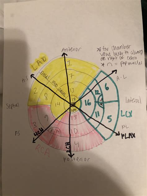
lv wall echo | wall motion chart echo lv wall echo Calculation of the left ventricular wall motion score index (WMSI) with transthoracic echocardiography allows the semi-quantification of left ventricular ejection .
If it is missing either one or both of these two things, it isn't real. In any case, the model number should start out with either a M or a N before following up with four or five numbers. If the model number turns up a different product, it .
0 · wall segments echo printable charts
1 · wall motion chart echo
2 · wall motion abnormalities echo
3 · lvh echo guidelines
4 · lvh echo criteria wall thickness
5 · lv wall thickness echo measurement
6 · lv wall thickness echo
7 · lv function assessment by echo
42 min read. Max Verstappen took the win in Las Vegas after overcoming a five-second penalty, a collision with George Russell and Ferrari’s Charles Leclerc to win the.Get up to speed with everything you need to know about the 2023 Las Vegas Grand Prix, which takes place over 50 laps of the 6.2-kilometre Las Vegas Strip Circuit in Nevada, USA, on Saturday, November 18. Using the links above you can find the full weekend schedule, including details of practice and qualifying sessions, support races, press .
Standardized myocardial segmentation and nomenclature for echocardiography. The left ventricle is divided into 17 segments for 2D echocardiography. One can identify these segments in .Wall motion is assessed in each segment of the left ventricle (Figure 1; refer to Segments of .Electronic calipers should be positioned on the interface between myocardial wall and cavity, and the interface between wall and pericardium. Perform at end-diastole (previously defined) .Wall motion is assessed in each segment of the left ventricle (Figure 1; refer to Segments of the Left Ventricle). Regional wall motion abnormalities are defined as regional abnormalities in .
Our LV calculator allows you to painlessly evaluate the left ventricular mass, left ventricular mass index (LVMI for the heart), and the relative wall thickness (RWT). Read on and discover all the details of our LV mass .
Calculation of the left ventricular wall motion score index (WMSI) with transthoracic echocardiography allows the semi-quantification of left ventricular ejection .Assessment of left ventricular systolic function has a central role in the evaluation of cardiac disease. Accurate assessment is essential to guide management and prognosis. Numerous echocardiographic techniques are used in the .
The first and most commonly used echocardiography method of LVM estimation is the linear method, which uses end-diastolic linear measurements of the interventricular . LV Function and Haemodynamic Assessment Echocardiography. SYSTOLIC FUNCTION. Global Function. stroke volume: end-diastolic volume – end-systolic volume; .
wall segments echo printable charts
LVM and RWT. LVM is the acronym for Left Ventricular Mass. LV mass (LVM) is a vital prognostic measurement we obtain with echocardiography to manage hypertension. RWT is the acronym for Relative Wall Thickness and is an . Each echocardiogram includes an evaluation of the LV dimensions, wall thicknesses and function. Good measurements are essential and may have implications for .Standardized myocardial segmentation and nomenclature for echocardiography. The left ventricle is divided into 17 segments for 2D echocardiography. One can identify these segments in multiple views. The basal part is divided into six segments of 60° each.
Electronic calipers should be positioned on the interface between myocardial wall and cavity, and the interface between wall and pericardium. Perform at end-diastole (previously defined) perpendicular to the long axis of the LV, at or immediately below the level of .
Wall motion is assessed in each segment of the left ventricle (Figure 1; refer to Segments of the Left Ventricle). Regional wall motion abnormalities are defined as regional abnormalities in contractile function. Ischemic heart disease is the most common cause of .

Our LV calculator allows you to painlessly evaluate the left ventricular mass, left ventricular mass index (LVMI for the heart), and the relative wall thickness (RWT). Read on and discover all the details of our LV mass calculator and its variables: Definitions of abnormal LV mass index; PWd normal range; and; IVSd in echo ️ Calculation of the left ventricular wall motion score index (WMSI) with transthoracic echocardiography allows the semi-quantification of left ventricular ejection fraction (LVEF).Assessment of left ventricular systolic function has a central role in the evaluation of cardiac disease. Accurate assessment is essential to guide management and prognosis. Numerous echocardiographic techniques are used in the assessment, each . The first and most commonly used echocardiography method of LVM estimation is the linear method, which uses end-diastolic linear measurements of the interventricular septum (IVSd), LV inferolateral wall thickness, and LV internal diameter derived from 2D-guided M-mode or direct 2D echocardiography. This method utilizes the Devereux and Reichek .
LV Function and Haemodynamic Assessment Echocardiography. SYSTOLIC FUNCTION. Global Function. stroke volume: end-diastolic volume – end-systolic volume; cardiac output: Q = SV X HR = (Aortic Area x V x Tej) x HR. Q = cardiac output Aortic area = cross sectional area V = velocity for each beat Tej = time period during ejection HR = heart rateLVM and RWT. LVM is the acronym for Left Ventricular Mass. LV mass (LVM) is a vital prognostic measurement we obtain with echocardiography to manage hypertension. RWT is the acronym for Relative Wall Thickness and is an additional reference value that can help further classify the . Each echocardiogram includes an evaluation of the LV dimensions, wall thicknesses and function. Good measurements are essential and may have implications for therapy. The LV dimensions must be measured when the end-diastolic and end-systolic valves (MV and AoV) are closed in the parasternal long axis (PLAX) view.Standardized myocardial segmentation and nomenclature for echocardiography. The left ventricle is divided into 17 segments for 2D echocardiography. One can identify these segments in multiple views. The basal part is divided into six segments of 60° each.
Electronic calipers should be positioned on the interface between myocardial wall and cavity, and the interface between wall and pericardium. Perform at end-diastole (previously defined) perpendicular to the long axis of the LV, at or immediately below the level of .Wall motion is assessed in each segment of the left ventricle (Figure 1; refer to Segments of the Left Ventricle). Regional wall motion abnormalities are defined as regional abnormalities in contractile function. Ischemic heart disease is the most common cause of . Our LV calculator allows you to painlessly evaluate the left ventricular mass, left ventricular mass index (LVMI for the heart), and the relative wall thickness (RWT). Read on and discover all the details of our LV mass calculator and its variables: Definitions of abnormal LV mass index; PWd normal range; and; IVSd in echo ️ Calculation of the left ventricular wall motion score index (WMSI) with transthoracic echocardiography allows the semi-quantification of left ventricular ejection fraction (LVEF).
Assessment of left ventricular systolic function has a central role in the evaluation of cardiac disease. Accurate assessment is essential to guide management and prognosis. Numerous echocardiographic techniques are used in the assessment, each .
The first and most commonly used echocardiography method of LVM estimation is the linear method, which uses end-diastolic linear measurements of the interventricular septum (IVSd), LV inferolateral wall thickness, and LV internal diameter derived from 2D-guided M-mode or direct 2D echocardiography. This method utilizes the Devereux and Reichek .
LV Function and Haemodynamic Assessment Echocardiography. SYSTOLIC FUNCTION. Global Function. stroke volume: end-diastolic volume – end-systolic volume; cardiac output: Q = SV X HR = (Aortic Area x V x Tej) x HR. Q = cardiac output Aortic area = cross sectional area V = velocity for each beat Tej = time period during ejection HR = heart rateLVM and RWT. LVM is the acronym for Left Ventricular Mass. LV mass (LVM) is a vital prognostic measurement we obtain with echocardiography to manage hypertension. RWT is the acronym for Relative Wall Thickness and is an additional reference value that can help further classify the .
best creed cologne 2022

best calvin klein coat for warmth
In this article, we’ll give you the best seven methods of spotting a fake Louis Vuitton Belt LV Initiales. So buckle up, cause we’re in for a ride! How to legit check Louis Vuitton Belt LV Initiales? The Logo Method. The Print Method. The Buckle Method. The Font Method. The LV Lettering Method. The Serial Number Method. The Box Method.
lv wall echo|wall motion chart echo


