lv end-diastolic dimensions should be measured at qrs | lv end diastolic lv end-diastolic dimensions should be measured at qrs LV end-diastolic dimensions should always be measured the upstroke of the QRS. Partition values allow you separate the left atrium from the left ventricle. LV measurements taken from low parasternal windows overestimate true values. The diagnosis of LV hypertrophy is . OCTA. Pirkt polisi. Pieteikt atlīdzību. Vairāk par OCTA apdrošināšanu. Veselības apdrošināšana. Saņemt piedāvājumu. Pieteikt atlīdzību. Vairāk par veselības apdrošināšanu. Ceļojumu apdrošināšana. Atlaide ergo.lv. -15% Pirkt polisi. Pieteikt atlīdzību. Vairāk par ceļojumu apdrošināšanu. Visi apdrošināšanas veidi. Pieteikt atlīdzību.
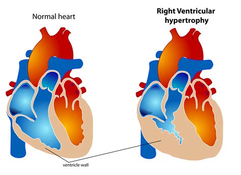
0 · right ventricular hypertrophy measurements
1 · lv heart size chart
2 · lv end diastolic measurements
3 · lv end diastolic
4 · left ventricular hypertrophy measurements
5 · left ventricular heart size chart
6 · left ventricular heart measurements
2 talking about this. Radio home for ESPN 1100AM, FOX Sports 1340AM, NBC Sports 920AM and.
LV end-diastolic dimensions should always be measured the upstroke of the QRS. Partition values allow you separate the left atrium from the left ventricle. LV measurements taken from low parasternal windows overestimate true values. The diagnosis of LV hypertrophy is .Perform at end-diastole (previously defined) perpendicular to the long axis of the LV, at or immediately below the level of the mitral valve leaflet tips. LV mass = 0.8x (1.04x .Aortic dimensions should be measured using 2D imaging from the PLAX window. Indices should be obtained using the inner-edge to inner-edge (IE-IE) methodology in end-diastole, defined .
Normal (reference) values for echocardiography, for all measurements, according to AHA, ACC and ESC, with calculators, reviews and e-book.To measure end-diastolic diameter use the section of the MMode in which the ventricle is largest, shortly before the walls begin to move inward (onset of the QRS complex). For the end . The LV dimensions must be measured when the end-diastolic and end-systolic valves (MV and AoV) are closed in the parasternal long axis (PLAX) view. The measurement .LA volume should be measured at left ventricular end systole (largest LA size at the frame just before mitral valve opening) using the Simpson’s biplane MoD and then indexed to BSA. Since .
QRS duration increased by 5.4 ms for every 100 g increase in LV mass, and by 4.6 ms for each 10 mm increase in LV end-diastolic diameter. The amplitude increased by 0.8 mm for every . In patients with LBBB, LV length (r = 0.32, p = 0.03), mass (r = 0.39, p = 0.01), diameter (r = 0.34, p = 0.02), and LV end-diastolic volume (r = 0.32, p = 0.04) had positive . LV end-diastolic dimensions should always be measured the upstroke of the QRS. Partition values allow you separate the left atrium from the left ventricle. LV measurements taken from low parasternal windows overestimate true values. The diagnosis of LV hypertrophy is based on wall thickness. 2005.I will review the fundamentals of the correct techniques for accurate LV measurement, explain the timing of end diastole/systole in regards to linear measurements, discuss caliper location and outline 6 pitfalls to avoid when measuring the left ventricular wall and chambers.
Perform at end-diastole (previously defined) perpendicular to the long axis of the LV, at or immediately below the level of the mitral valve leaflet tips. LV mass = 0.8x (1.04x [(IVS+LVID+PWT) 3 -LVID 3 ] + 0.6 gramsAortic dimensions should be measured using 2D imaging from the PLAX window. Indices should be obtained using the inner-edge to inner-edge (IE-IE) methodology in end-diastole, defined as the onset of the QRS complex. All values should be indexed to height and not BSA.
Normal (reference) values for echocardiography, for all measurements, according to AHA, ACC and ESC, with calculators, reviews and e-book.To measure end-diastolic diameter use the section of the MMode in which the ventricle is largest, shortly before the walls begin to move inward (onset of the QRS complex). For the end-systolic dimension, pick the region in which the ventricular cavity is smallest. The LV dimensions must be measured when the end-diastolic and end-systolic valves (MV and AoV) are closed in the parasternal long axis (PLAX) view. The measurement is performed in the basal portion of the LV by the chordae. Left ventricular dimensions. Left ventricular geometry and mass. References.
LA volume should be measured at left ventricular end systole (largest LA size at the frame just before mitral valve opening) using the Simpson’s biplane MoD and then indexed to BSA. Since apical views that are optimised for the LV will foreshorten the LA, dedicated apical 4-chamber and 2-chamber images should be acquired to maximise LA .QRS duration increased by 5.4 ms for every 100 g increase in LV mass, and by 4.6 ms for each 10 mm increase in LV end-diastolic diameter. The amplitude increased by 0.8 mm for every 100 g increase in LV mass. In patients with LBBB, LV length (r = 0.32, p = 0.03), mass (r = 0.39, p = 0.01), diameter (r = 0.34, p = 0.02), and LV end-diastolic volume (r = 0.32, p = 0.04) had positive correlations with QRSd.
LV end-diastolic dimensions should always be measured the upstroke of the QRS. Partition values allow you separate the left atrium from the left ventricle. LV measurements taken from low parasternal windows overestimate true values. The diagnosis of LV hypertrophy is based on wall thickness. 2005.
I will review the fundamentals of the correct techniques for accurate LV measurement, explain the timing of end diastole/systole in regards to linear measurements, discuss caliper location and outline 6 pitfalls to avoid when measuring the left ventricular wall and chambers.Perform at end-diastole (previously defined) perpendicular to the long axis of the LV, at or immediately below the level of the mitral valve leaflet tips. LV mass = 0.8x (1.04x [(IVS+LVID+PWT) 3 -LVID 3 ] + 0.6 grams
Aortic dimensions should be measured using 2D imaging from the PLAX window. Indices should be obtained using the inner-edge to inner-edge (IE-IE) methodology in end-diastole, defined as the onset of the QRS complex. All values should be indexed to height and not BSA.
Normal (reference) values for echocardiography, for all measurements, according to AHA, ACC and ESC, with calculators, reviews and e-book.To measure end-diastolic diameter use the section of the MMode in which the ventricle is largest, shortly before the walls begin to move inward (onset of the QRS complex). For the end-systolic dimension, pick the region in which the ventricular cavity is smallest. The LV dimensions must be measured when the end-diastolic and end-systolic valves (MV and AoV) are closed in the parasternal long axis (PLAX) view. The measurement is performed in the basal portion of the LV by the chordae. Left ventricular dimensions. Left ventricular geometry and mass. References.
order more links for michael kors watch
LA volume should be measured at left ventricular end systole (largest LA size at the frame just before mitral valve opening) using the Simpson’s biplane MoD and then indexed to BSA. Since apical views that are optimised for the LV will foreshorten the LA, dedicated apical 4-chamber and 2-chamber images should be acquired to maximise LA .QRS duration increased by 5.4 ms for every 100 g increase in LV mass, and by 4.6 ms for each 10 mm increase in LV end-diastolic diameter. The amplitude increased by 0.8 mm for every 100 g increase in LV mass.

right ventricular hypertrophy measurements
lv heart size chart
For your convenience, we combined best escape rooms in Latvia in one place. Choose and make a booking for any quest room you like and don’t forget to leave a feedback after the game. Best price guarantee!
lv end-diastolic dimensions should be measured at qrs|lv end diastolic


